Pterygium

A pterygium is a triangular growth of abnormal conjunctival tissue that invades the cornea.

Its development is not related to injury or inflammation. 90% of pterygia are located nasally, the nose side of the eye. Both eyes are frequently involved but often only one.

The pterygium can advance to involve the visual axis causing marked loss of visual acuity.

The pterygium can be distinguished from a pingueculum, which is a small-elevated yellowish mass, which can become inflamed. The pterygium grows in the interpalpebral fissure (that is the exposed part of the eye) as an elevated fleshy mass of the conjunctiva.

Symptoms

Burning, irritation, tearing and a foreign body sensation may accompany the growth of a pterygium onto a cornea. Astigmatism may be induced and may lead to decreased vision. As the apex of the pterygium approaches the visual axis, glare and reduced contrast sensitivity appear.

For poorly understood reasons the pterygium growth may stop at any stage. It may remain so for the rest of the patient’s life or at a later date again start growing.

Etiology and Epidemiology

There is a worldwide distribution of pterygium but it is more common in warm, dry climates. Studies have shown a strong relationship between ultraviolet radiation and the formation of pterygia.

A study in Australia identified a number of risk factors:

  • Living in the tropics
  • Working in a sandy outdoor environment
  • Not wearing sunglasses
  • Not wearing a hat

Local drying of the conjunctiva and tearfilm abnormalities may also be contributing factors.

The highest incidents occur between the ages of 20-49 and rarely occur under the age of 15.

Treatment

The decision to remove a pterygium is dependent on the patient’s symptoms, complications caused by the pterygium and the interest in cosmetic improvement. Recurrences may be more frequent in young adults. Virtually all post-operative recurrences occur within the first year after surgery and often within 6-8 weeks. With recurrence there is a higher incidence of growth into the visual axis.

Medical Treatment

Eye drops such as preservative free lubricants, vasoconstrictors and mild corticosteroids can relive the symptoms. Some believe ultraviolet-blocking spectacles can prevent progression.

Surgical

The pterygium is removed surgically and the method depends on the surgeon doing the operation.

  1. The pterygium is avulsed.
  2. The pterygium is excised and the underlined sclera is left exposed, that is bare sclera.
  3. Transplantation of the head of the pterygium away from the cornea beneath the superior edge of the adjacent normal conjunctiva.
  4. Keratectomy, is when a superficial layer of the cornea is removed during the pterygium operation on the cornea.  The pterygium may then be removed, and the sclera left bare as above or the head of the pterygium can be transplanted.

Adjunctive Therapy

A number of therapies are used to decrease the risk of recurrence after surgical removal of the pterygium. The treatment used again depends on the surgeon’s preference. Each has attractive features, but none is without drawbacks.

The following may be used:

  1. Steroid drops.
  2. Thiotepa, a radiomimetic agent, which presumably obliterates proliferation of vascular cells and is very rarely used today.
  3. Mitomycen C is an antineoplastic-antibiotic agent, which inhibits the synthesis of proteins.
  4. Beta-radiation. Strontium-90 applicators are used to radiate the site of the pterygium after surgical removal.
  5. Performing an auto-graft in which a piece of conjunctiva from the same eye is glued or sutured onto the site that the pterygium was removed.

Future Prospects

The first report of a surgical treatment of a pterygium is more than 3000 years old. The management of pterygia and recurrent pterygia is improving yet many questions remain unanswered. Future studies may elucidate the cause of the pterygium as well as the cause of complications related to the adjunctive therapy after pterygium surgery and prevent recurrence.


The role played by Cornea transplant co-ordinator

Many hundreds of corneal grafts taking place annually in South Africa, the need for which continues to increase.

Corneal transplantation, also known as corneal grafting, is a surgical procedure where a damaged or diseased cornea is replaced by donated corneal tissue (the graft). When the entire cornea is replaced it is known as penetrating keratoplasty and when only part of the cornea is replaced it is known as lamellar keratoplasty.

What is recoded below is a snapshot of the process undertaken to ensure that the donor cornea is harvested and delivered to the surgeon timeously for the graft to take place.

The graft co-ordinator is contacted by a hospital to notify them of the passing of a donor patient. The medical history of the donor patient is reviewed to confirm that the  donor cornea is suitable. Anyone can become an organ donor . Persons under the age of 18 require parental permission. The exclusions are as follows: positive virology results for infectious diseases such as HIV/AIDS, Hepatitis B or C, septicaemia, previous ocular tumours and those in which possible COVID- 19 has not been excluded.

The co-ordinator has approximately 12 hours to harvest the cornea. It is often a difficult time to approach the family and the co-ordinator needs to be sensitive when discussing donation with the family, where organ donation has not been previously consented to.

Donated tissue is procured by trained personnel who work for the Eye Bank and this takes place at the hospital or funeral home. The process of recovering the tissue takes approximately 30 minutes. Great care is taken to preserve the appearance of the donor. The tissue recovery does not compromise funeral arrangements.

The tissue is carefully stored in preservation media and transported to the Eye Bank where it is placed in a temperature regulated fridge. The tissue is evaluated for infections and imperfections before being issued. Less than 1% of corneas previously donated to the Eye Bank have been deemed unsuitable for transplant. In the unlikely event of this occurring, the Eye Bank has a policy regarding the medical discarding of tissue through an established hospital procedure.

A corneal transplant is usually performed within 1 to 10 days after donation, depending on the completion of testing, the location of the receiving hospital and the preparation of the selected recipient.

The gift of sight is usually made anonymously but the recipient family can write a letter of thanks to donor family.


Presbyopia

What is Presbyopia?

Are you always searching for your reading glasses? Have you ever wished that there was a solution to this problem? When you are in front of the television your glasses are either in the bedroom, kitchen, car or even at the office. With the development of technology there is an operation available which could eliminate this problem and make the “old” eye as good as new.

Reading problems will start to occur at the age of about 40 years and affects just about everybody after 45. When the eyes are focusing close up, the lens changes its shape. The diameter increases and light rays are focused on the retina. As we get older the lens grows and when the space is too limited to adjust its shape, reading problems start to occur.

Modern technology has proved that by increasing the diameter of the eye it is possible to eliminate problems with reading. This enables the lens to change shape and focus again close up for reading.

Increasing the diameter is achieved by inserting four plastic arches into the white of the eye(sclera). These arches have a very specific design and are being produced, under license, in America.

The recovery process depends on the age of the patient. People under the age of 50 will recover quicker than those over 50. Recovery can take up to six months and requires a lot of exercise. This can be compared to a broken arm or leg: When the plaster of paris is removed the bone is thin and the muscles are weak. The muscles need to regain their strength as soon as there is sufficient space available for the lens to change its shape while reading.

Who qualifies for this operation?

Patients over the age of 40.

No underlying refraction errors. Other refractive procedures need to be treated (e.g. LASIK operation) before the reading problems can be addressed. Patients with no underlying eye diseases. Some systematic illnesses may be a relative contra-indication (e.g. Diabetes)

How is the operation performed?

The implants are done under general anaesthetic and the patient goes home that same day. The patient will have to come for a one-day, one week, one month and a three-month follow-up after the operation. Only one eye is done at a time. In some cases it will only be necessary to do one eye. Some patients will be able to function effectively if the dominant eye is done first.


What are the dangers involved?

As with any other operation there is always the risk of infection. To prevent this the operation is performed under sterile conditions in theatre. It is possible that the arches may move which will reduce the effectiveness of the procedure. This can be corrected by repositioning the arches under anaesthetic. In a small percentage of cases the eye has reject the arches. This is rare but is possible.

What can I expect immediately after the operation?

  • For the first week the eye will be red and scratchy.
  • Vision may fluctuate, especially for reading.
  • Headaches may follow due to the eyedrops prescribed.
  • Nausea due to the stretching of the eye. The body must adapt to the new shape.

Contact your local ophthalmologist for more information.


What is Glaucoma?

Glaucoma

What is glaucoma?

In the normal eye a watery liquid is constantly produced and drained away through tiny drainage canals. This fluid maintains a pressure within the eye that remains constant as long as the fluid drains as quickly as it is produced. The normal intraocular pressure is between 10 and 21 mm Hg. Glaucoma may occur when there is a build-up of pressure within the eye due to clogged or covered drainage canals, where the eye fluid cannot drain away.

Glaucoma is a physical condition within the eye and it is not a form of cancer or tumor, it is not infectious or contagious, it is not a danger to life itself. Glaucoma is a painless and silent disease and if it is left untreated or neglected, it will result in loss of vision and even lead to blindness.

Are there different types of glaucoma?

Yes, there are many different types, but there are two major types: Chronic and Acute. Chronic open angle glaucoma (COAG) is the most common form glaucoma. In COAG the entrance to the drainage canals is open, but there is a clogging problem inside the drainage canals. The disease is slow, causes gradual loss of small patches of vision, it is a lifelong condition and responds well to medication and surgery.

Acute closed-angle glaucoma is rare. The eye pressure goes up quickly because the drainage canals are suddenly blocked. This causes severe headaches or eye pain, nausea, rainbows around light and severely blurred vision. This glaucoma demands immediate medical attention, day or night.

Does glaucoma run in families?

Yes, if one parent has COAG the chances of offsprings are 10 time greater than in the non glaucoma population.

How is glaucoma treated?

Generally speaking, glaucoma cannot be cured, but it can be controlled. Once diagnosed, it requires constant, lifelong care. The major goal of glaucoma treatment is to keep the eye pressure in a range that prevents further glaucoma damage. This can be done with eye drops, pills, laser treatments and microsurgery.

What advice is there for patients with glaucoma?

Follow your eye specialist’s instructions, use medications regularly, know what your medications are, know their side effects, tell your doctors that you have glaucoma and what medications you are using, call your eye specialist about any unusual changes in your eyes or vision, plan regular check-ups on time.


World Down Syndrome Day

FOR THIS ARTICLE, NDSS INTERVIEWED DANIELLE LEDOUX, MD ASSISTANT IN OPHTHALMOLOGY AT CHILDREN’S HOSPITAL, BOSTON AND INSTRUCTOR IN OPHTHALMOLOGY AT HARVARD MEDICAL SCHOOL

Down syndrome has effects on the developing eye which can impact the proper development of vision. Eye disease is reported in over half of patients with Down Syndrome, from less severe problems such as tear duct abnormalities to vision threatening diagnoses such as the early onset of cataracts. Particular attention should be paid to vision in people with Down syndrome.

What is Different About the Eyes in Individuals with Down Syndrome?

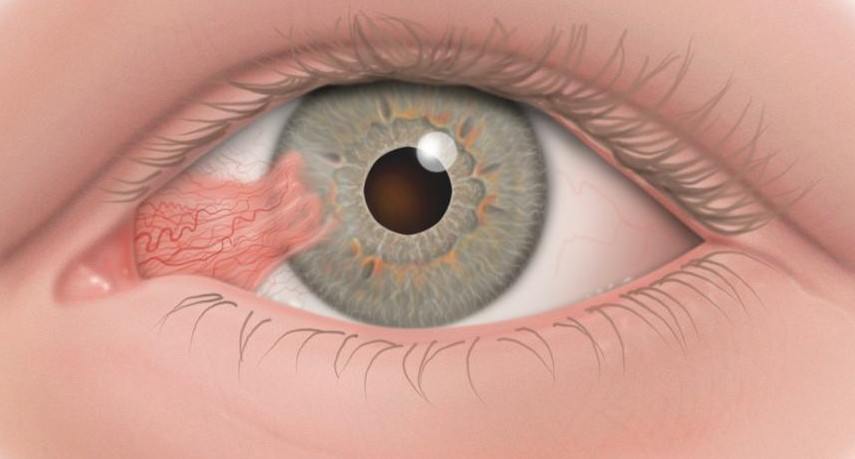
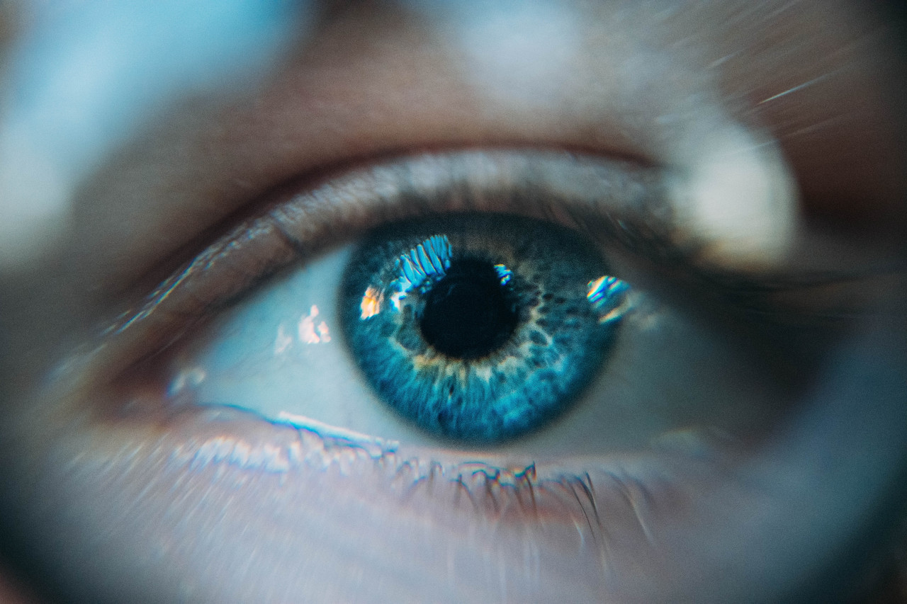
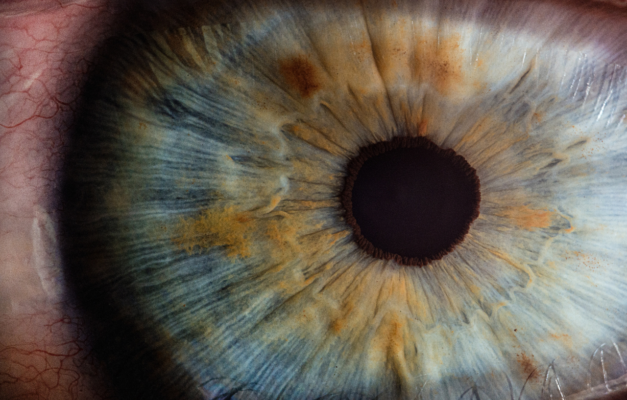
Dr. Ledoux: As any family member of a person with Down syndrome knows, there are characteristic features about the eyes. This includes upward slanting of the eyelids, prominent folds of skin between the eye and the nose, and small white spots present on the iris (the coloured part of the eye) called Brushfield’s spots. These spots are harmless.

Do Most Children with Down Syndrome Need Glasses?

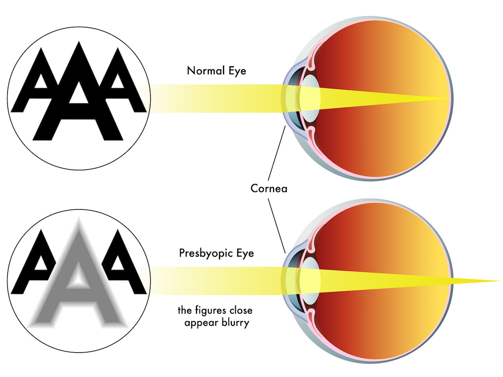
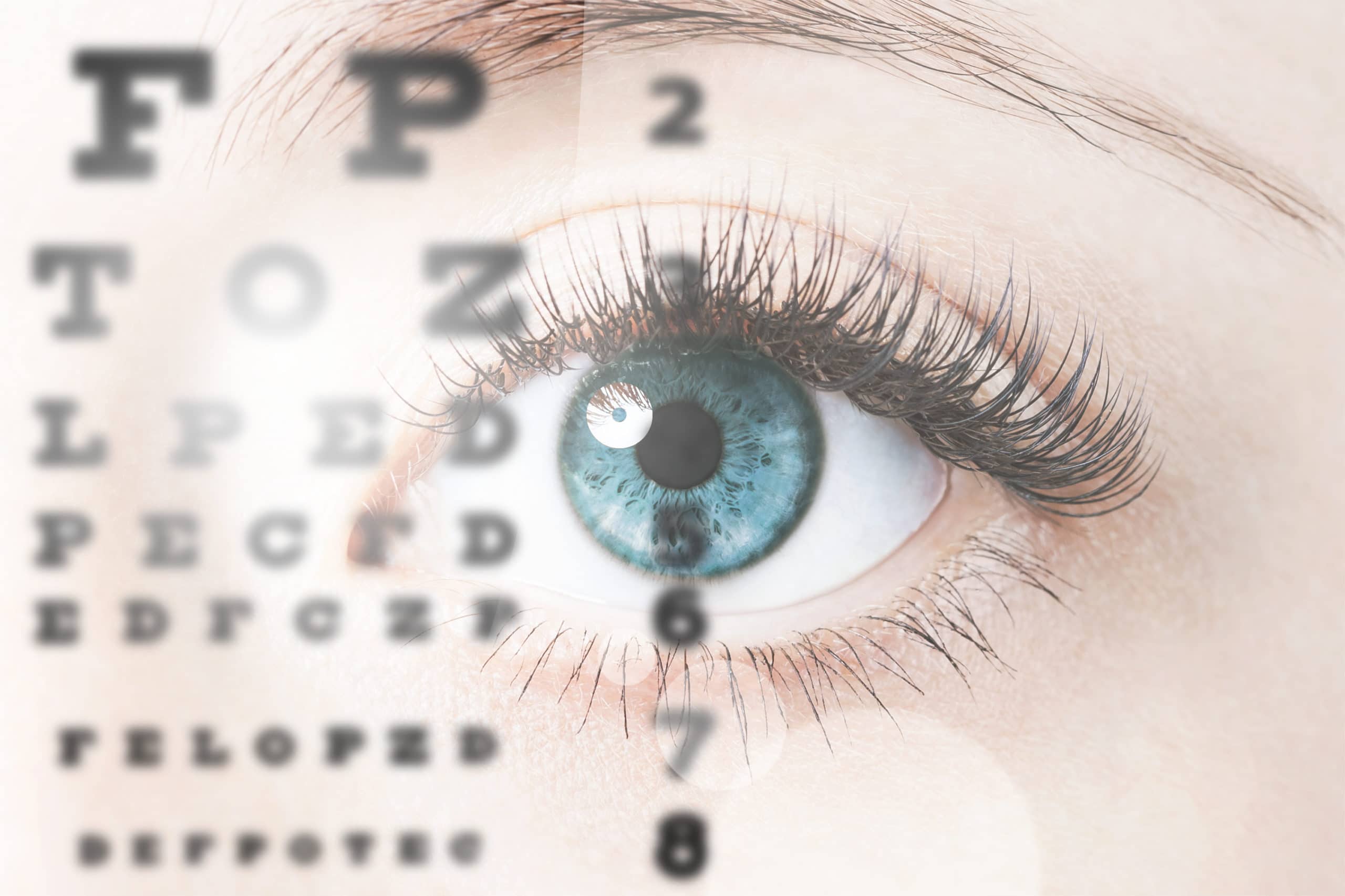
Dr. Ledoux: Refractive error (the need for glasses) is much more common in children with Down syndrome than in the general population. This refractive error can be hyperopia (farsightedness), astigmatism, or myopia (nearsightedness). Another problem is weak accommodation (difficulty changing the focusing power of the eye from distance to near). We can test this easily in the office, and if detected, we will prescribe glasses that have bifocals. Some of my patients have difficulty adjusting to glasses, but once they get accustomed to having the glasses on their face, their vision is significantly better and often their eye alignment improves as well.

What Are Common, but Less Serious, Eye Abnormalities Affecting Patients with Down Syndrome?

Dr. Ledoux: In addition to the need for eyeglasses, many children with Down syndrome have tear duct abnormalities. Family members will notice this as frequent discharge and tearing from the eyes, worsened by colds. We generally recommend firm massage over the space and down towards the nose (tear sac region) 2-3 times a day to attempt to open the tear duct. If this continues beyond 1-2 years of age, the tear ducts may need to be opened by a surgical procedure.

Strabismus (eye misalignment/ squint) is also more common. Family members may notice that the eyes do not line up well with each other, but often the strabismus can be subtle, even to the pediatrician. The folds of skin I mentioned between the eyes and the nose can also cover up the underlying strabismus, or make the eyes appear as if they are crossing even if they are not. It is important to diagnose strabismus as a child, as crossed eyes can result in amblyopia (loss of vision also known as lazy eye) and loss of stereopsis (the use of the two eyes together or depth perception).

How Can the Strabismus Be Treated?

Dr. Ledoux: Sometimes, simply glasses alone are enough to straighten eyes with strabismus. If glasses are needed, we always start there. If the eyes continue to have strabismus despite the correct pair of eyeglasses, then we recommend strabismus surgery (eye muscle surgery). This is a one to two hour procedure. Unfortunately, our patients with Down syndrome are more likely to require more than one surgery to align their eyes as they don’t always respond as predictably to strabismus surgery as the general population with strabismus would.

What Are the More Severe Eye Problems That Might Develop?

Dr. Ledoux: My greatest concern is congenital cataracts (lack of clearness to the lens of the eye). If visually significant cataracts are present early in a child’s eye, then a clear image is not delivered to the brain and therefore the brain can never “learn” to see. This is a severe form of amblyopia known as form deprivation amblyopia. While we can take our time removing a cataract in an adult patient, if significant cataracts are present very early in a child’s life, and not removed, can result in lifelong poor vision. In that situation, even if the cataract is removed when the child is older, the vision never improves significantly. This is what makes early detection of cataracts in infants and children so important. A child with Down syndrome will be evaluated by the paediatrician at birth, and referred to an ophthalmologist if something abnormal is detected. There is also a unique form of cataract in Down syndrome patients that we have found in our research. However, depending on how developmentally delayed the person is, they may not be able to communicate that they can’t see. For this reason, I recommend any patient with Down syndrome, no matter what age, have a complete eye examination if they are starting to show reduced cognitive function, or changes in their normal activities.

Are There Other Eye Conditions in Down Syndrome That Can Cause Loss of Eyesight?

Dr. Ledoux: I mentioned amblyopia (commonly called “lazy eye” which is decreased vision) which can be caused by multiple different eye problems such as strabismus, severe ptosis (eyelid droop), cataracts, or even uncorrected refractive error, especially if one eye needs a much stronger eyeglass prescription than the other. Ptosis is usually easier to appreciate but strabismus and significant refractive error can be very difficult for the paediatrician to diagnose. There are other more rare problems which can occur with the optic nerve or retina of the eye which can sometimes cause vision loss and unfortunately are generally not treatable. Nystagmus (a rhythmic shaking of the eyes) can also occur.

What is the Recommended Eye Care for Children with Down Syndrome?

Dr. Ledoux: The American Academy of Paediatrics (AAP) and the Down Syndrome Medical Interest Group (DSMIG) recommend evaluation of the red reflex of the eyes at birth to look for cataracts, as well as to assess the eyes for strabismus or nystagmus. The red reflex is essentially the “red eye” seen in photographs, which is the normal reflex of the retina when struck by direct light. If the eyes don’t look normal, then the infant will be referred to a paediatric ophthalmologist – a physician who has completed specialty training in medical and surgical management of the child’s eye. We, along with the AAP and the DSMIG, recommend a child with Down syndrome has their first eye exam by an ophthalmologist experienced in patients with special disabilities (for example, a paediatric ophthalmologist) by six months of age. After that, children with Down syndrome, even if they are without symptoms, should see an ophthalmologist every one to two years. If any eye problems are detected, they will be followed more frequently.

What Sort of Symptoms Might We See If a Child Has an Eye Problem?

Dr. Ledoux: Unfortunately, children with Down Syndrome often do not complain about their eye problems, either because they don’t notice the problem or because they can’t communicate the problem well enough. Signs to look for include squinting or closing one eye shut, an unusual head tilt, crossing or wandering of one or both eyes, or light sensitivity. In some severe cases, the sign of vision problems may be a regression in overall function or loss of developmental milestones. Ptosis will be seen as a lid droop, and a blocked tear duct will result in daily tearing and discharge.

Any Thoughts for Parents of a Child With Down Syndrome Who Are Concerned About the Eye or Vision?

Dr. Ledoux: Getting regular eye exams is very important in children with Down syndrome because eye disorders are so common and are difficult for the paediatrician to diagnose. Because the examination can be difficult for both the child and the doctor, it is best to have the examination done by an ophthalmologist skilled in dealing with children with developmental delays. Don’t be surprised to find out your child needs glasses; if needed, the glasses will help the vision, and possibly the eye alignment, as well as to help in the development of normal vision pathways in the brain. This will help with your child’s learning and functioning.


THE COVID-19 PROCEDURE WHEN VISITING THE JHB EYE HOSPITAL

THE COVID-19 PROCEDURE WHEN VISITING THE JHB EYE HOSPITAL
Overview:
The global pandemic of the coronavirus disease 2019 (COVID-19) was first reported on the 31st December 2019 by the World Health Organization following a cluster of pneumonia cases in Wuhan City, Hubei Province of China. Severe acute respiratory syndrome coronavirus 2 (SARS-CoV-2) has been confirmed as the causative virus of COVID-19. To date, COVID-19 has become a global pandemic and it is continuing to spread across the globe. COVID-19 affects different people in different ways. Most infected people will develop mild to moderate illness and recover without hospitalization. For more information on the virus please refer to the following websites: https://www.nicd.ac.za/ and https://covid19.who.int/.
Our Procedures:
Should a patient require eye surgery following a consultation with an ophthalmic surgeon, they will be requested to visit one of the laboratories in their area to have a COVID-19 test done prior to their procedure. This test should be performed no more than 3-5 days prior to their admission to the Johannesburg Eye Hospital. Should the test results be positive, the scheduled procedure will be postponed to a future date. A negative result will allow the procedure to go ahead as scheduled. Should the patient be a minor or person with a disability or an elderly person then the person accompanying this patient would also need to have a COVID-19 test done with a negative result prior to the patients admission. Between the date of testing and date of surgery the patients are requested to have minimal social contact and if possible to isolate at home.
Prior to a patient’s admission, they will be requested by the receptionist at the ophthalmic surgeon’s practice to complete the JHB EYE Hospital’s pre-authorisation form and anaesthetist form. This can be found on our website at https://www.jhbeye.com/patient…/covid-19-policy/ and may be emailed it to the hospital staff at [email protected].
Patients and accompanying visitor’s will be required to wear face masks for the duration of their visit to the JHB Eye Hospital and will be required to have a health screening done at Reception upon arrival. Patients are also reminded to practice safe social distancing at all times.
Unfortunately, no visitors are allowed into the Eye ward, Laser centre or Medwedge Stepdown Facility currently.
We thank you for your understanding and co-operation during this time. The health and safety of our staff and our patients is our utmost priority.

Eye Safety

Eye Safety during the festive season
Dr Irfaan Hasrod MBBCh(Wits), FCOphth(SA), MMed Ophth(Wits), DipOpht(SA)
The holiday season is a time spent relaxing and creating memories with our loved ones. It is also a time when we see an increase in trauma to patients’ eyes. Here is a quick safety check list to keep your eyes safe and preserve the valuable gift of sight:
1.Always wear safety glasses when doing any DIY chores at home, this will prevent foreign bodies entering the eye, as well as preventing devastating globe lacerations.
2.Think twice before purchasing any sharp-edged toys, projectile toys, and laser toys for the little ones. They are always culprits in ocular trauma seen in children during the holidays.
3.Champagne cork injuries to the eyes can be avoided by using a towel when opening champagne bottles.
4.Use UV protected eyewear when enjoying the beautiful outdoors. Excess UV exposure can cause photokeratitis (a form of sunburn to the eyes).
5.Do not delay in seeking professional help in the event of any injury/visual disturbances/eye pain you may have. If any eye problem is diagnosed and treated early the outcome is usually superior.
For more information please visit https://www.jhbeye.com/

Staff wellness

Johannesburg Eye Hospital Wellness day 18/12/2020
Nursing 101 taught me to: Promote health, prevent illness, preserve life and alleviate suffering…
I am proud to say that in 2020 the management of Johannesburg Eye Hospital took this principle to heart.
Health care personnel, have the tendency to neglect their own health to take care of the needs their patients. However, during our wellness day, we focused on the wellbeing of the health care personnel.
We conducted the following test:
• weight, height and BMI with a consultation form our dietitian
• blood pressure, blood glucose, blood cholesterol, ECG, HIV
testing and counselling and Hepatitis antibody testing and immunization
• consultation from our medical doctor
• eye screening and a consultation from our ophthalmologist
Thank you is such a small word, but it is said with a warm and grateful heart to all the staff at the Johannesburg Eye Hospital. Your hard work, commitment and endurance during a very tough year are greatly appreciated. You are an amazing team. However, a great team is a reflection of awesome leadership. Thank you to our management and stakeholders for the inspiration, guidance and support.
I wish you and your family peace, health and happiness during this festive season.